Sign In

  • Forgot your password?
  • Need a new account?

Register


Spinning Disk Confocal Microscope | SIMTRUM Photonics

Spinning Disk Confocal Microscope

Spinning disk confocal microscopy is an innovative fluorescence imaging technique that overcomes the limitations of traditional point-scanning confocal microscopy in terms of imaging speed and phototoxicity, making it particularly suitable for observing living cells and rapid dynamic processes. Its core lies in a high-speed rotating disk, which is densely populated with tens of thousands of tiny pinhole arrays. When laser light passes through these pinholes, it simultaneously generates multiple focused spots on the sample, enabling multipoint parallel scanning. This parallelism is the key to its speed advantage.

Multipoint parallel scanning greatly enhances imaging speed, reaching up to hundreds of frames per second, allowing scientists to capture rapidly changing events within cells. At the same time, because each spot receives less optical energy per unit time, it significantly reduces photodamage to living samples and photobleaching of fluorescent dyes, making it an ideal choice for long-term, low-phototoxicity live-cell imaging.

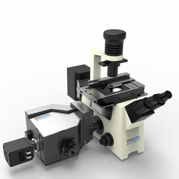
SIMSCOP's spinning disk series combines excellent imaging performance, dynamic imaging friendliness, portable hardware integration, and a rich software operating system, making it suitable for various research and industrial-grade imaging applications.

1.png


Product Core Specifications

  • High-speed spinning disk: 15,000 rpm
  • Multi-wavelength laser: Supports multi-channel lasers from 400 nm to 750 nm
  • Large field of view: Up to 25 mm
  • High-speed acquisition: 100 fps @ 2048×2048 pixels
  • Software functions: Large image stitching, 3D imaging rendering, time-lapse imaging, Z-stack scanning
2.png


Customizable Spinning Disk

With an ultra-high rotation speed of 15,000 RPM, our spinning disk ensures complete and uniform scanning of the entire field of view even at minimal exposure times, producing smoother images free of artifacts. The extreme rotational speed substantially shortens the laser exposure time per point per unit time, thereby minimizing the cumulative light energy absorbed by fluorophores. Moreover, we provide customizable spinning disk options with variable pinhole pitch and diameters to match specific user requirements.

3.png


Excellent Imaging Performance

This system is built upon an advanced spinning disk confocal optical architecture and optimized light path design, delivering high-resolution, large-field-of-view, and high-uniformity imaging while maintaining high-speed performance. Compared with traditional widefield and point-scanning confocal systems, it demonstrates significant advantages in image clarity, background suppression, and field-of-view utilization, making it capable of meeting multi-level demands ranging from fine structural observation to large-scale sample analysis.


14.jpeg

Higher Resolution

With high numerical aperture (NA) objectives and optimized optical design, the system achieves an XY resolution of 230 nm and a Z-axis optical section thickness of 600 nm, making subcellular structures and minute details clearly visible. Compared with traditional confocal or widefield microscopes, this system offers significant advantages in signal-to-noise ratio and image contrast, enabling the detection of weak fluorescent signals and supporting high-precision quantitative analysis and structural studies.

Expansive Field of View

With a maximum field of view of up to 25 mm × 25 mm, the system enables comprehensive sample coverage in a single capture, significantly reducing the time spent on repositioning between views. When paired with the software's large-image stitching capability, it facilitates seamless panoramic visualization—from subcellular features to whole-mount tissue architecture. Relative to traditional microscopes, this system offers enhanced large-area scanning efficiency, while preserving sharpness and edge-to-edge fidelity with minimal optical distortion.

图片翻译 (12).jpeg
图片翻译 (13).jpeg

More Uniform Illumination

We have carried out optimized designs tailored separately for laser conditions and LED light fields. Through laser homogenization and speckle suppression techniques, local overexposure and optical artifacts are greatly reduced. Compared with conventional laser scanning microscopes, this system ensures high precision and reliability in quantitative analysis while maintaining high resolution, and significantly reduces the risks of phototoxicity and photobleaching, providing robust support for long-term live-cell imaging.

Designed for Dynamic Imaging

Thoroughly optimized for live-cell and biological specimen imaging, this system strikes the perfect balance between high-speed data acquisition and minimal sample damage. Whether you are tracking fast cellular dynamics or performing extended time-lapse studies, it consistently delivers high-quality, reproducible image data for reliable and comparable results. When set against traditional microscopy systems, it stands out with clear advantages in acquisition speed, image stability, and sample compatibility.


7.png

High-Speed Acquisition (100 fps @ 2048×2048 pixels)

The system supports an acquisition speed of up to 100 fps, enabling the capture of rapid cellular movements and transient changes in microscopic structures. Combined with a wide field of view and optimized detector design, this performance far exceeds that of traditional confocal microscopes, while maintaining high signal-to-noise ratio and image contrast. High-speed acquisition ensures that rich information is obtained within a short period, making complex dynamic processes clearly visible and providing more comprehensive data support for scientific research.

Capturing Dynamic Processes

By integrating time-series acquisition with intelligent image processing capabilities, the system allows real-time documentation of key biological events, including cell division, migration, and signaling responses. Its low-light illumination strategy, combined with an optimized optical design, preserves sample viability over extended experiments, preventing phototoxicity or photobleaching from repeated excitation. In contrast to conventional live-cell imaging systems, this solution offers prolonged continuous observation windows and markedly enhanced reliability and reproducibility of experimental outcomes.

8.png
9.png

Sample-Friendly Imaging

By employing laser homogenization and meticulous light intensity regulation, the system effectively prevents localized overexposure. Optimized excitation wavelengths and a low-illumination approach minimize sample damage, preserving cellular health and stability throughout extended experiments. When paired with high-speed acquisition and real-time dynamic capture, this system empowers researchers to obtain rapid, high-quality imaging results without sacrificing sample viability—offering an ideal balance between performance and biological compatibility.


User-Friendly Hardware Operation

With a meticulously optimized hardware architecture, this system delivers an exceptional balance of flexibility, expandability, and imaging performance. Its modular design, coupled with seamless multi-mode switching, enables users to tailor the system configuration to specific experimental requirements—all while maintaining superior image quality and minimizing the system footprint. This provides a robust and versatile platform for a wide range of research applications.


Modular Design

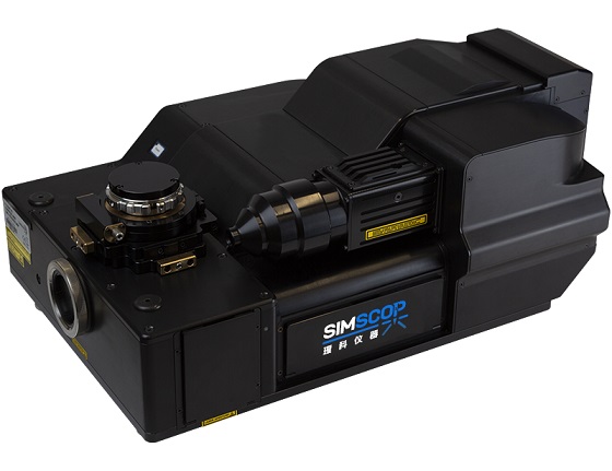
The system adopts a compact and highly configurable spinning disk module, delivering unparalleled flexibility and expandability for optical platforms. This modular design significantly reduces the system's footprint on the optical bench without sacrificing any imaging quality. Its diverse configuration options are capable of meeting your multiple requirements for imaging performance and system integration, allowing users to freely configure the system according to experimental needs without compromising image quality or occupying excessive space, thus providing a reliable foundation for a wide variety of experiments.


1.png


Fast Multi-Mode Switching

The system supports rapid switching between widefield and confocal modes, offering both manual and motorized options. Through integrated design, users can switch between different imaging modes almost seamlessly, ensuring both fast large-field observation and immediate access to confocal mode when high resolution is required. Compared with conventional microscopes that require disassembly and reassembly or lengthy adjustments when switching modes, this system's fast switching improves experimental efficiency while maintaining stable imaging performance, providing seamless support for multi-protocol experimental workflows.


2.png


Spinning Disk Confocal Microscope Specifications

Module Parameter SimDISK-Standard SimDISK-Pro SimDISK-U
Spinning
Disk
Module
Disk Speed 1000 – 15000 RPM
Scan Rate 100 – 1000 Fps
Disk Geometry
(Diameter/Spacing)

Standard: Single-pattern disk: 50/250 slit, suitable for high-throughput laser applications;

Optional: Multi-pattern disk: 50/250 slit, 60/220 slit, 35/350 slit, 65/165 slit, 50/350 slit, suitable for high-throughput laser, LED illumination, high-resolution laser, and ultra-high-throughput laser applications

Filter Wheel Emission filter wheel: 8-position (25mm), includes 4 single-band filters Excitation filter wheel: 8-position (25mm), includes 4 single-band filters, multiband filters can be added
Emission filter wheel: 8-position (25mm), includes 4 single-band filters, multiband filters can be added
Includes one 405nm narrow-band filter
Widefield & Spinning Disk Switching Manual Motorized Manual
Beam Conditioning Homogenizer + Square Beam Adapter
Excitation Light Interface Standard: SMA905 interface, 400 μm core multi-mode fiber, 0.22 NA
Customizable: FC/PC, FC/APC interfaces, and other core diameters of multi-mode fibers
FC/APC interface, single-mode polarization-maintaining fiber for 405nm laser
Field of View (FOV) Max. 28 mm diameter
Disk
Configuration
Camera Port C-mount sCMOS camera (compatible with CCD/EMCCD camera) × 1 port C-mount sCMOS camera (compatible with CCD/EMCCD camera) × 2 ports C-mount sCMOS camera (compatible with CCD/EMCCD camera) × 1 port
Dimensions 251 × 251 × 380 mm 251 × 251 × 380 mm /
Laser
Module
Laser Source 1. 405/488/532/640nm multi-mode laser (power per wavelength >500mW, continuous laser spectral bandwidth <5nm, power stability <2%, SMA905 interface, 400 μm core multi-mode fiber, independent control per wavelength, power adjustment accuracy: 1%, integrated electro-optical unit)
2. 405/488/561/640nm multi-mode laser (output power per wavelength >500mW, continuous laser spectral bandwidth <5nm, power stability <2%, SMA905 interface, 400 μm core multi-mode fiber, independent control per wavelength, power adjustment accuracy: 1%, integrated electro-optical unit)
3. Optional despeckle module (recommended)
Includes standard 405nm single-mode polarization-maintaining laser (wavelength 405±5nm, output power 45mW, stability <±1.5%, TTL modulation, FC/APC interface, power adjustment accuracy: 1%, integrated electro-optical unit)
Detector
Module
Camera Type C-mount sCMOS Camera
Camera Specifications 95%@600nm Peak QE, 6.5 μm × 6.5 μm Pixel Size, 2048 × 2048 Resolution, 13.3mm × 13.3mm Effective Area, Noise: 0.2e−, Full-Well Capacity 45ke−, Air and Water Cooling Compatible
Microscope
Frame &
Body
Microscope 1. Split System (Fully Automated Inverted Fluorescence Microscope):
• 10W LED, 100W Mercury Lamp, 130W Metal Halide Fluorescence Light Source LUMOS X, LED Fluorescence Light Source LUMOS3, LED Fluorescence Light Source LUMOS7
• FN25 Eyepiece 10x/25mm, 360° rotatable viewing tube, interpupillary distance adjustment 48~75mm, beam splitting ratio 100:0, 50:50, 0:100 (PX55 FS8 PLUS supports motorized switching), optional tilting binocular tube
• Coded / Motorized 8-position fluorescence turret
• Fluorescence filter block (DAPI / 395), fluorescence filter set (AT-EGFP / FITC / Cy2 / AlexaFluor 488), fluorescence filter set (AT-TRITC / Cy3 / TagRFP / AlexaFluor 546) (customizable upon request)
2. All-in-One System
1. All-in-One System:
• Reflected light source: 12V/100W long-life halogen lamp
• 5-position nosepiece with DIC slot, motorized 5-position nosepiece with DIC slot (optional)
Objectives Uplan APO 4x/0.16 W.D.=13mm, CG=0.17
Uplan APO 10x/0.45 W.D.=3.1mm, CG=0.17
Uplan APO 20x/0.80 W.D.=0.6mm, CG=0.17
Uplan APO 40x/0.95 W.D.=0.18mm, CG=0.17
Uplan APO 40x/0.95 W.D.=0.18mm, CG=0.11-0.23
Uplan APO 60x/1.42 Oil. W.D.=0.18mm, CG=0.17
Uplan APO 100x/1.45 Oil. W.D.=0.13mm, CG=0.17
Plan S-APO 5X/0.15
Plan S-APO 10X/0.3
Plan S-APO 20X/0.45
Plan S-APO 50X/0.8
Plan S-APO 100X/0.9 (Optional)
Motorized Stage XY Stepper motor XY microscope stage: Travel range 120mm (X) × 80mm (Y), Max. speed 30mm/s, Resolution <1 μm, Unidirectional repeatability ≤1 μm, Repeatability ≤2.5 μm, Max. load 1 kg
Motorized Stage Z Motorized Z-axis, Travel range 13mm (Up 9mm + Down 4mm), Stepper motor, Min. resolution 0.01 μm, Repeatability ±0.5 μm, Max. speed 6.4mm/s Piezo Z-stage, Travel range 500 μm, Closed-loop resolution 13.5 nm, Repeatability 0.05% F.S., Load capacity 2 kg
Software
Control
Software Functions System control, multi-color fluorescence localization processing, Z-stack processing, large image stitching, image analysis, imaging data management, 3D image rendering, deconvolution, etc. System control, Z-stack processing, large image stitching, image analysis, imaging data management, 3D image rendering, deconvolution, etc.

FISH Fluorescence In Situ Hybridization and Spinning disk confocal microscope system

FISH(Fluorescence In Situ Hybridization)is an important way to locate a specific nucleic acid sequence in a cell or tissue. By labeling DNA or RNA probes, combined with fluorescence microscopy, FISH technology can accurately locate and detect specific genes, chromosome structures and functional regions at the cellular level, which has important application value for cytogenetics, oncology, developmental biology and other fields.


However, due to the limitation of the optical diffraction limit, the imaging resolution of the FISH probe is about 300 nm when the excitation light with a wavelength of 561 nm is used. When the distance between two or more fluorescent probes in the nucleus is less than the imaging resolution, it will not be able to accurately count them. To achieve accurate statistics of HER2 gene levels in positive patients, higher resolution imaging techniques are needed. In addition, wide-field fluorescence microscopy is not suitable for imaging tissue sections because it detects too much defocusing blur.

Observation mode Advantages Disadvantages
Wide field fluorescence microscope One of the main imaging tools for 
the pathological diagnosis of FISH.
Low resolution for the optical diffraction limit.
Relatively simple to use, low cost. It uncapable of counting fluorescent probes 
whose nuclear distance is less than the resolution.
The coordinates of FISH points can be 
easily determined in cells.
Not applicable to imaging tissue sections.
Laser scan confocal microscope The imaging signal-to-noise ratio is improved and the imaging quality is better. The imaging speed is slow and not suitable for 
practical FISH diagnostic applications.
Multiple synaptic structural markers can be 
visualized simultaneously by fluorescent immunohistochemistr.
The quantum efficiency of using 
photomultiplier tubes is relatively low.
Spinning disk confocal microscope Faster imaging speed and higher imaging resolution. Spatial resolution is a little lower than 
point scan confocal microscope.
It is an excellent tool for quantifying the number of 
multiple synaptic components simultaneously and for estimating protein levels in tissue slices.
Faster and better 3D imaging.
sCMOS cameras have high quantum efficiency and high imaging quality.
Reduce photobleaching and phototoxicity.


By combining SpinDisk Standard with FISH technology, our system breaks the limitations of traditional imaging and provides a superior solution for FISH imaging with the following key features:

  • Large-field image stitching: Easily stitch multiple local images into a complete image, significantly expanding the field of view.
  • Multi-channel imaging and fusion: Supports simultaneous capture of multiple fluorescent channels and fusion of them to provide more comprehensive detection results.
  • Image processing and analysis: Built-in powerful image processing software to optimize image quality, improve contrast and sharpness. Provides quantitative analysis tools to easily calculate the intensity, distribution and correlation of fluorescence signals.
  • 3D reconstruction and rendering: Realize 3D reconstruction of the sample, provide realistic 3D imaging effect, and help in-depth understanding of the spatial structure of the sample.
  • Accurate fluorescence positioning: For multi-color fluorescent labels, the system can accurately locate and analyze the distribution of signals.
  • Z-stack data processing: Acquire and overlay images along the Z-axis to reconstruct the three-dimensional shape of the sample.
  • Automatic multi-dimensional scanning: supports multi-dimensional automatic scanning to achieve full coverage and efficient data acquisition.

This fully integrated imaging solution enables users to obtain clearer and more accurate results in FISH experiments, providing strong support for scientific research and clinical applications.


SpinDisk Confocal Microscopy Acquistion Atlas


High-Definition Visual Assets: Should you require high-definition authentic images and video footage, we invite you to get in touch with us.

Mouse nerve 3D tomography, 20X objective lens, scanning depth ~100 microns

(Original Image)


Mouse nerve tomography 3D reconstruction, 20X objective lens, scanning depth ~100 microns

Mouse nerve 3D tomographic scanning, 60X objective lens, scanning depth ~100 microns

(Original Image)


Mouse nerve tomography 3D reconstruction, 60X objective lens, scanning depth ~100 microns

(The picture has been processed with background noise reduction)

Zebrafish 3D tomography (Microscope system uses Olympus IX83)


Cell mitosis CETN1-GFP green, SiR-DNA blue (Microscope system uses Nikon Ti2)

BPAE-60X1.2NA-405nm-CF

BPAE-60X1.2NA-488nm-CF

BPAE-60X1.2NA-525nm-CF

BPAE-60X1.2NA-Polychromatic fusion-CF

SpinDisk Standard-20X -0.5NA-405/488/525
Multi-color fusion and large image Mosaic of mouse intestinal tissue embedded section samples - full image size 1.8mmX1.25mm composed of 56 partial images


Mouse intestinal tissue embedding section-60X-1.2NA-405nm-WF


Mouse intestinal tissue embedding section-1.2NA-405nm-CF


Mouse intestinal tissue embedding section-60X-1.2NA-488nm-CF


Mouse intestinal tissue embedding section-60X-1.2NA-Bicolor fusion-CF

Mouse cranial nerve, 20X05NA, 488nm laser channel
0.5um layer cut 100um thickness 3D reconstruction


SIMSCOP Spinning Disk Confocal Core Components Test Report

This test report presents measured verification of the core optical components of the SIMSCOP spinning disk confocal microscope, covering micron-scale pinhole fabrication morphology and pitch calibration, the effective illumination spot size coverage on the disk surface, and laser homogenizer energy distribution uniformity testing.

1. Custom Spinning Disk Spot Size and Homogenization Efficiency Test

A beam shaping homogenizer combined with a square spot conversion module was used to perform multiple tests on the effective illumination area of the excitation beam on the spinning disk target surface and the gray value profile along the diagonal energy cross-section [cite: 3]. The measured spot can fully cover the 22–28mm disk field of view, and the homogenization efficiency remains stable above 87%–90%, effectively eliminating the over-bright center of the Gaussian beam and the dark corner edge effect.

Measurement Group 01: Diagonal cross-section spot and gray level uniformity analysis (Homogenization efficiency: 87.84%)
Diagonal spot measurement image 1
Measurement Group 02: Diagonal cross-section spot and gray level uniformity analysis (Homogenization efficiency: 89.58%)
Diagonal spot measurement image 2
Measurement Group 03: Diagonal cross-section spot and gray level uniformity analysis (Homogenization efficiency: 90.35%)
Diagonal spot measurement image 3
Test Conclusion: Disk surface spot size: 22 – 28 mm | Spot diagonal profile homogenization efficiency: 87.83% – 90.35%

2. Actual Measurement of Spinning Disk Pinhole Shape and Size

A high-magnification microscopic imaging system was used to perform geometric calibration and optical edge calibration of the micro/nano fabrication quality of the spinning disk pinhole array. By extracting the transmitted light gray level transition curve of the pinhole cross-section, the true aperture value, pitch consistency, and hole roundness are precisely measured, ensuring that confocal imaging has a high signal-to-noise ratio and ideal optical sectioning thickness.

Pinhole morphology micrograph 1
Pinhole morphology micrograph 2
Pinhole morphology micrograph 3

Spinning Disk Pinhole Actual Photo (Light Transmission Array Distribution)

In the light transmission test of the spinning disk micro-hole array, the overall pinholes are round and smooth, regularly distributed without adhesion, and no burrs, edge peeling, or defect obstruction were observed, ensuring high consistency of the excitation spot across the full field of view and high fluorescence excitation efficiency.

Spinning disk pinhole light transmission actual photo 1
Spinning disk pinhole light transmission actual photo 2

SIMSCOP Spinning Disk Confocal Microscope Real Acquisition Gallery and Application Cases

This real acquisition gallery presents a comparison of fluorescence microscopy imaging of biological tissue sections, focusing on the imaging quality, signal-to-noise ratio, and out-of-focus background suppression capability of the custom spinning disk under different pinhole diameters and spacings, as well as a real acquisition comparison with imported mainstream spinning disk modules.

1. Custom Spinning Disk Testing with Different Pinhole Parameters

Test results of the custom spinning disk with different pinhole (disk) parameters: The figure systematically compares the fluorescence imaging performance of different pinhole diameter and spacing combinations (65/165 μm, 65/245 μm, 50/250 μm, 35/350 μm, and 35/175 μm). The upper row shows the original field of view, and the lower row shows magnified views of the dashed box regions, used to compare local structural details and background suppression capability.
From the test results, it can be seen that different pinhole parameters have a significant impact on image contrast, background suppression, and signal intensity. Larger pinholes (such as 65 μm) can acquire higher fluorescence signals, but the background is relatively higher, and the local detail contrast is slightly reduced; after reducing the pinhole size to 35 μm, the out-of-focus background is significantly suppressed, and tissue boundaries and cellular structures become clearer, but the overall signal intensity is somewhat reduced. On the other hand, at the same pinhole size, increasing the pinhole spacing (such as 165 μm → 245 μm or 175 μm → 350 μm) can further reduce crosstalk between adjacent pinholes and improve image contrast, but excessively large spacing reduces sampling density, affecting illumination uniformity and imaging efficiency.
Considering imaging brightness, background suppression, and structural detail performance, different parameter combinations each have their advantages. Among them, 35/175 μm achieves a good balance between signal intensity and optical sectioning capability; 35/350 μm has the best background suppression effect, with overall lower brightness, but based on this spinning disk confocal microscope's high signal-to-noise ratio imaging system architecture, images can be reconstructed by adjusting the relative signal intensity (colormap rescaled); while the 65 μm pinhole scheme is more suitable for application scenarios requiring higher signal intensity.

65/165 um disk 65/245 um disk 50/250 um disk
65/165 um original field of view
65/245 um original field of view
50/250 um original field of view
35/350 um disk (with colormap rescaling) 35/175 um disk
35/350 um original field of view and colormap rescaling
35/175 um original field of view

2. Custom & Imported Spinning Disk Module Comparison

Under the same excitation power, exposure acquisition time, and detection-end configuration, the actual captured image quality of imported mainstream spinning disks and the custom Simscop spinning disk is compared. Under the common 50/250 μm pinhole specification, both show consistent high brightness and field lens flatness; while under high-resolution large-spacing modes (35/350 μm and 35/175 μm), the Simscop spinning disk exhibits superior optical sectioning suppression and high-contrast performance for cell boundaries and deep fine structures.

Test Group 01: Standard 50/250 μm Pinhole Specification Comparison
Imported disk, pinhole 50/250 μm
Imported disk pinhole 50/250um
Simscop disk, pinhole 50/250 μm
Simscop disk pinhole 50/250um
Test Group 02: Cell Structure Region Magnification and Background Crosstalk Comparison
Imported disk, pinhole 50/250 μm (global field of view)
Imported disk global and selected region
Simscop disk, pinhole 35/350 μm (left), 35/175 μm (right)
Simscop disk global and selected region
Test Conclusion: The Simscop custom spinning disk not only matches imported brands in image quality under high-throughput specifications (50/250 μm), but also supports customized multi-sector combinations, providing more targeted pinhole configuration solutions for thick samples and weak fluorescence scenarios.

* Images are only some cases and are affected by printing/display compression, so they may not achieve the best display quality. For more real acquisition cases and original images, please refer to the official website or consult a sales representative.

www.simscop.com

We are here for you!


Drop us an email at info@simtrum.com to get the Best Price today.

Want more technical information? Click Here for the professional Customized System/ Solutions.


Don't have time to search the products one by one? No worries. you can download the full range of SIMTRUM Product Line Cards.

Click it now.


Want to stay closer to the Market Dynamics and Technological 

Developments? Just take 5 seconds to Sign In as a member of 

SIMTRUM, we will bring you the most up-to-date news. 

(Sign in button on the top right of the screen).


Search Reset
Compare Model Drawings & Specs Availability Reference Price
(USD)
SpinDisk Basic Spinning Disk Confocal Microscope
Spinning Disk Confocal Microscope, SpinDisk Basic Series, Image frame rate 100fps@2048x2048, Resolution ~230nm, image depth <200um, No. of Laser - 4, Wavelength choice 405/445/488/525/561/640nm, sCMOS Camera detectors, Inverted or upright microscope, XYZ motorized stages, high image contrast, customized option
6-10 Weeks Request for quote
SpinDisk Advance
Spinning Disk Confocal Microscope, SpinDisk Advance Series, Image frame rate 100fps@2048x2048, Resolution ~230nm, image depth <200um, No. of Laser - 4, Wavelength choice 405/445/470/520/528/555/640nm, Dual sCMOS Camera detectors (single sCOMS optional), Inverted or upright microscope, XYZ motorized stages, Motorized filter electronics system, high image contrast, upgrade to SpinDisk SIM 100nm resolution, customized option
6-10 Weeks Request for quote

SpinDisk Advance - Parameter

SpinDisk Basic Spinning Disk Confocal Microscope - Parameter

SpinDisk Advance - Download

SpinDisk Basic Spinning Disk Confocal Microscope - Download

Accessories

Compare Model Drawings & Specs Availability Reference Price
(USD)